Which vascular condition is most commonly associated with a wandering spleen?
Which sonographic finding distinguishes focal nodular hyperplasia from hepatic adenoma?
Which structure is located between the fundus of the stomach and the diaphragm?
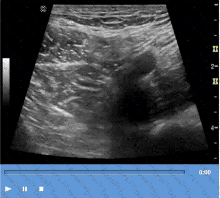
Which condition presents sonographically as an anechoic mass between the umbilicus and the bladder?
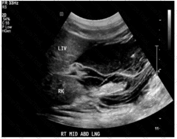
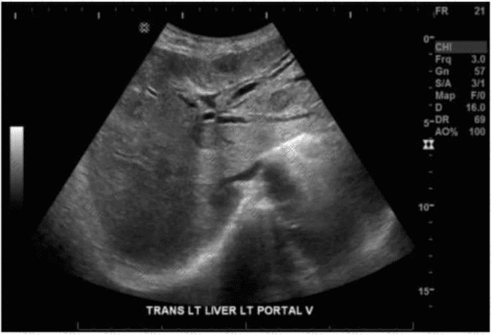
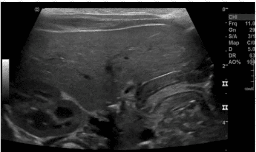
In which segment is the solid mass located in this transverse image of the liver?
Which action should a sonographer take if the abdominal aorta measures 5.5 centimeters in the anteroposterior diameter?
Which sonographic finding is associated with normal postprocedural Doppler of a transjugular intrahepatic portosystemic shunt (TIPS)?
What is the most common ultrasound appearance of the pancreas in mild acute pancreatitis?
A 60-year-old man presents to the emergency room, complaining of tearing pain in the chest and abdomen. Blood pressure readings from the two arms show a difference of more than 20 mm. Which ultrasound finding is most likely associated with this presentation?
Which sonographic finding indicates the need for immediate surgical intervention following testicular trauma?
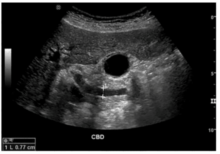
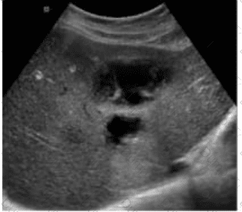
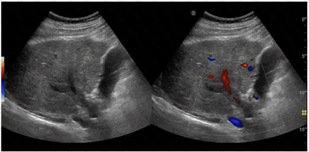
Which sonographic appearance of the bile ducts is demonstrated in this image?
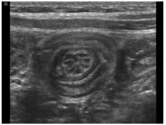
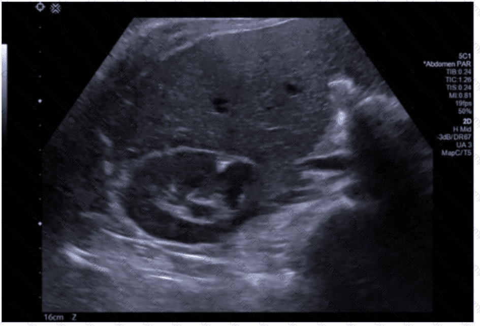
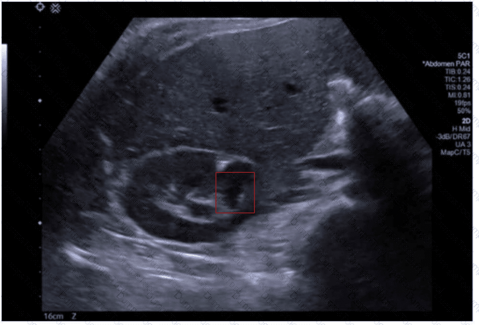
Identify the congenital anomaly.
Using your mouse, place the cursor on the appropriate region of the image and then left-click the mouse button to indicate your selection.
A patient presents with ampulla of Vater obstruction, distention of the gallbladder, and painless jaundice. Which condition is most likely associated with these findings?
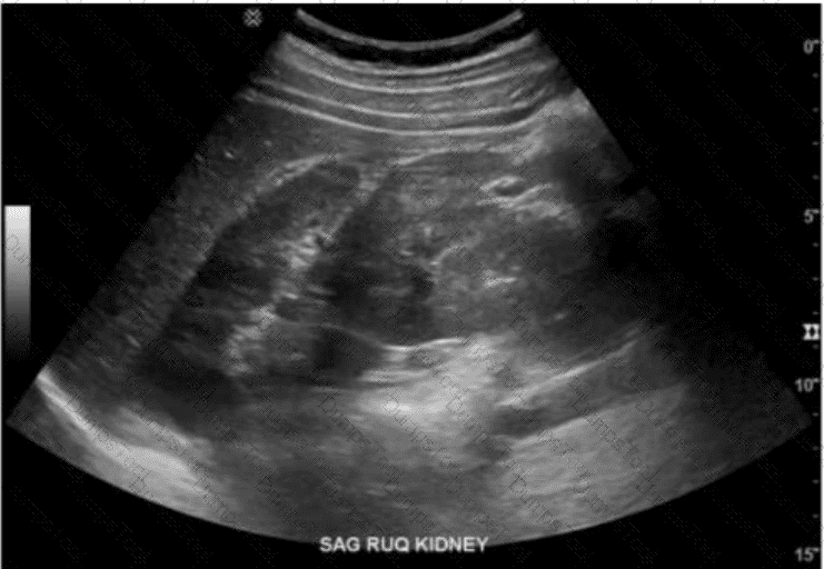
Which finding is an indication for renal biopsy to assess for renal failure?
Elevation of alpha-fetoprotein levels is a characteristic finding in which tumor?
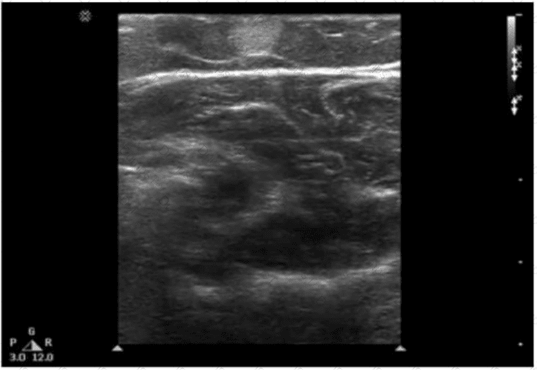
Which description is associated with the normal sonographic appearance of a tendon?
Which condition is most likely associated with this image of the common bile duct?
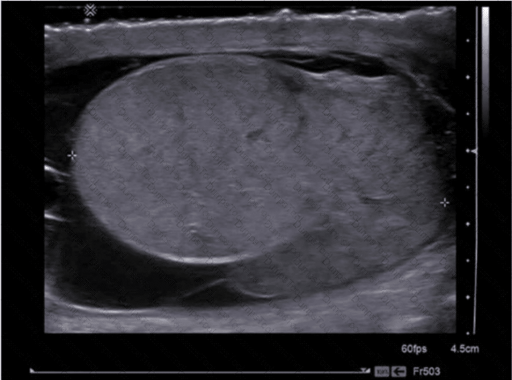
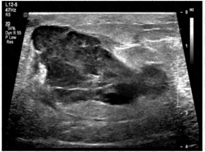
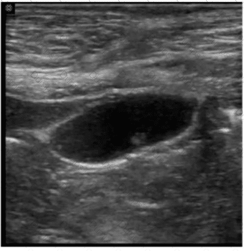
Which diagnosis is most consistent with this image from a patient with acute scrotal pain?
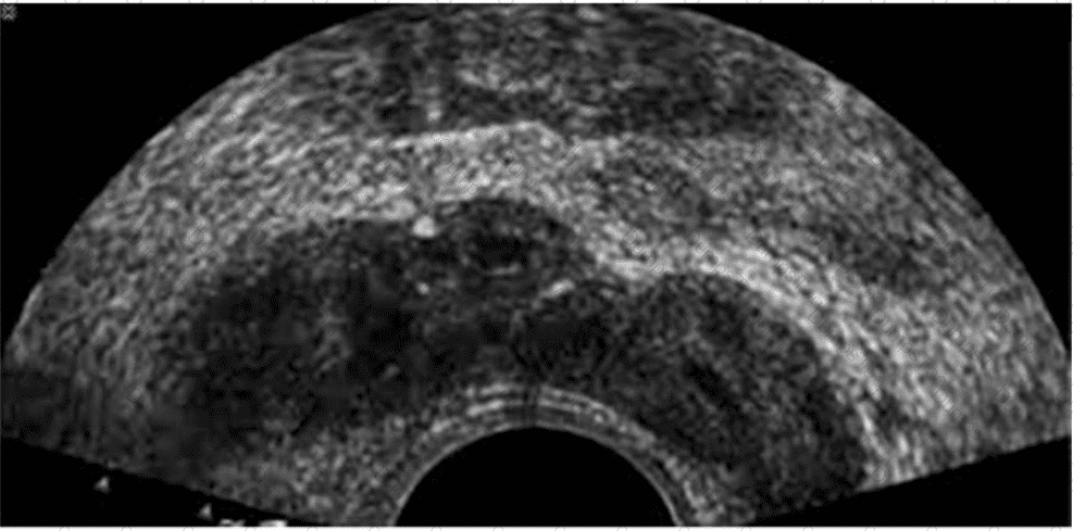
Which anatomical area of the male reproductive system is demonstrated in this endorectal image?
Which condition is most likely in a patient presenting with weight loss and fatigue along with elevated liver enzymes, elevated potassium, and decreased sodium?
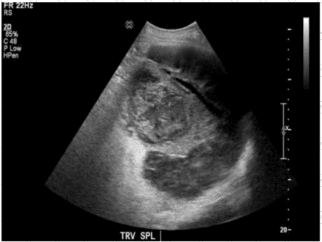
Which clinical finding is most likely associated with the splenic pathology demonstrated in this image?
Which common congenital anomaly is typically seen as a cystic midline anterior neck structure?
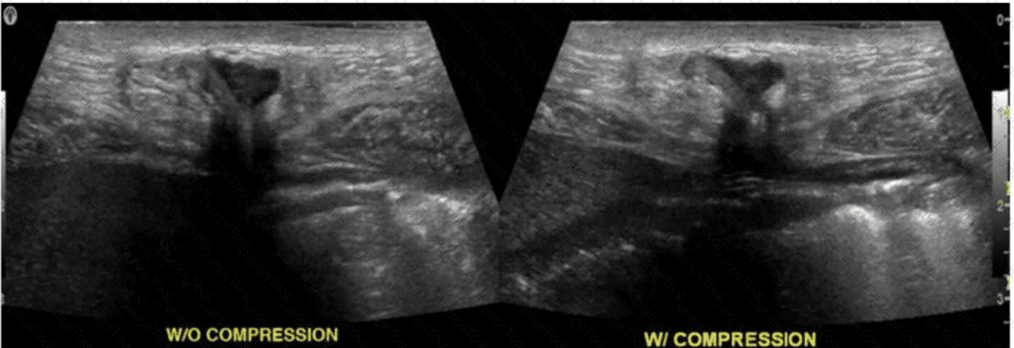
Which condition is most consistent with the sonographic appearance in this image of the abdominal wall?
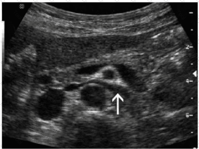
Which organ is drained via the vessel indicated by the arrow in this image?
Which finding is most likely demonstrated in this abdominal wall image of a patient with a history of atrial fibrillation?
Which abnormality is depicted in this image of a patient who presents with a fever following a liver biopsy?
The absence of which sonographic finding indicates the acute process depicted in these images?
Which technique would best assist the sonographer to verify the finding in this image obtained from the right upper quadrant?

TESTED 10 Jul 2026
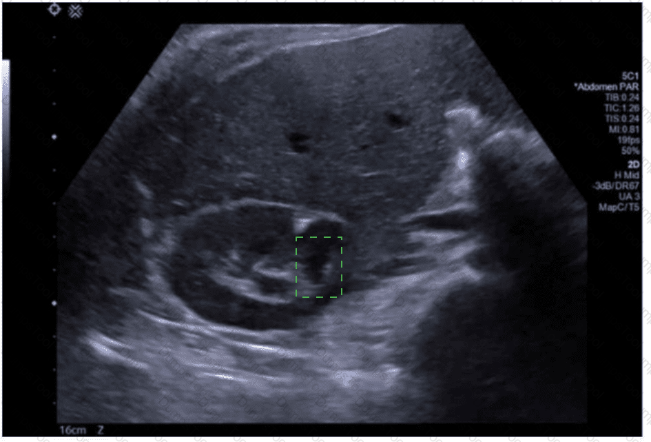
 An ultrasound of a fetus
AI-generated content may be incorrect.
An ultrasound of a fetus
AI-generated content may be incorrect.